
VeinViewer® for Research & Academic Institutions
Real-Time NIR Vascular Visualization for Clinical Research, Academic Medicine, and Evidence-Based Practice
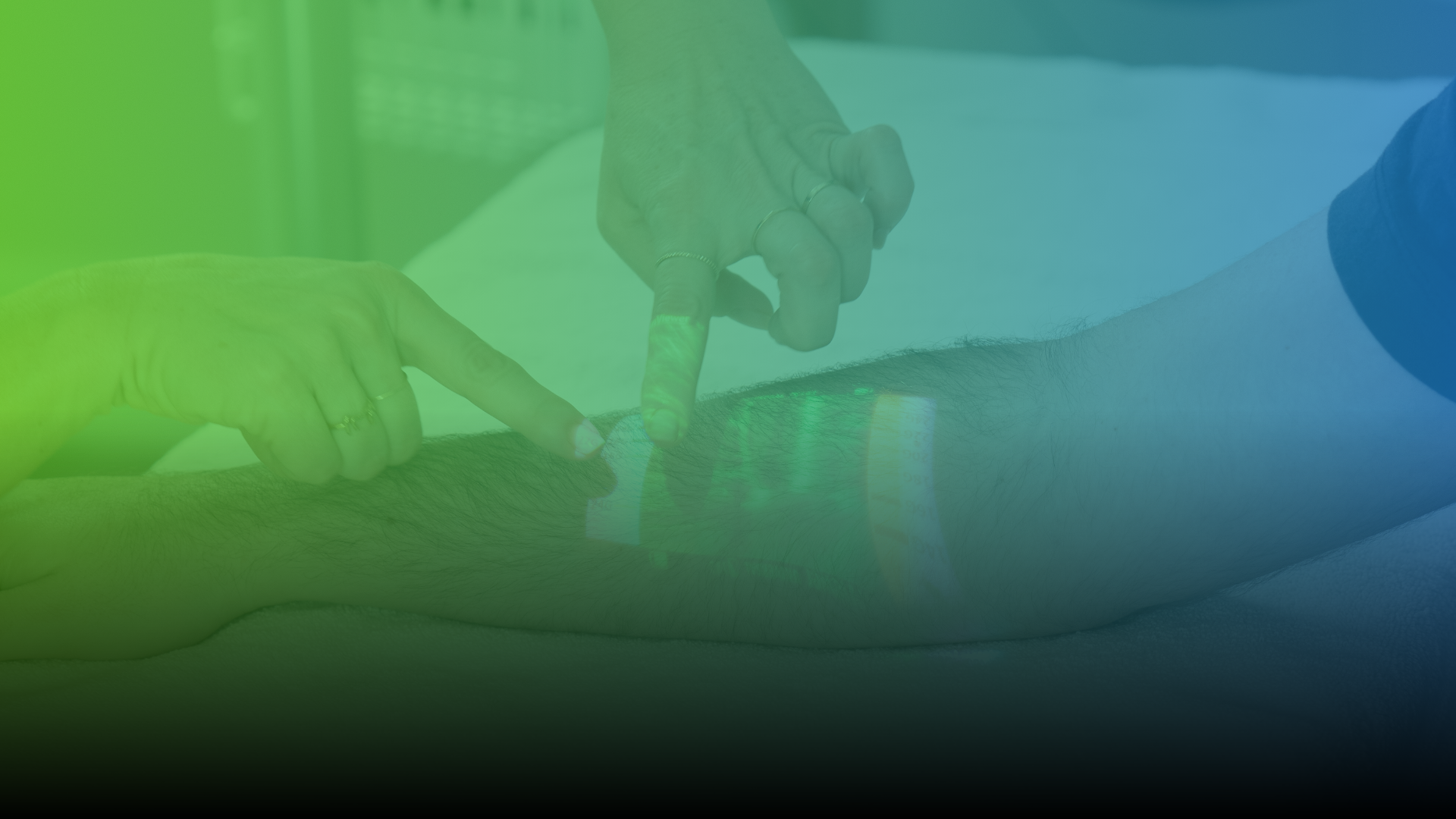
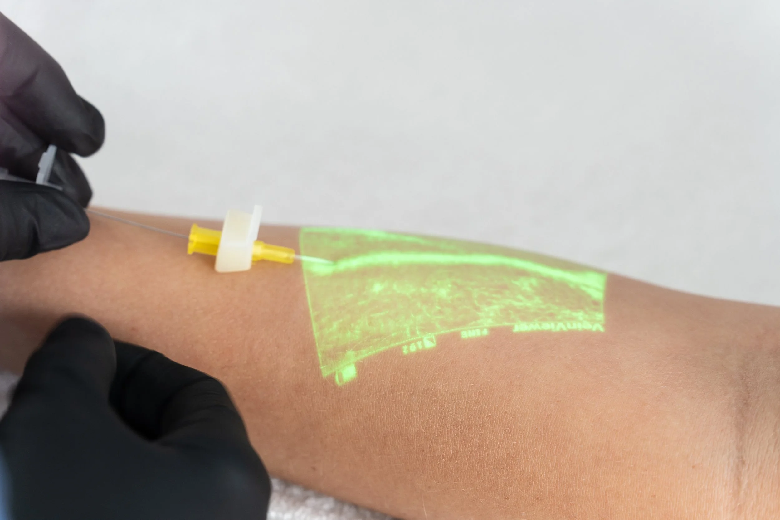
VeinViewer® near-infrared (NIR) technology projects a real-time, high-definition image of peripheral vasculature directly onto the skin — no gels, dyes, or disposables required. For researchers and academic clinicians, that means a repeatable, standardized vascular visualization tool that can support study design, procedural consistency, and outcome measurement across a wide range of clinical applications.
Whether you’re studying venous access in difficult populations, exploring the role of vein visualization in nursing education, or evaluating real-time imaging tools in veterinary or translational research, VeinViewer® Flex and VeinViewer® Vision2 are built to support your work.
Why Researchers Choose VeinViewer®
Variability in peripheral access technique is a known confounder in vascular access research. VeinViewer® provides a consistent imaging platform across providers and sites, helping research teams:
Standardize vein site selection methodology
Reduce operator-to-operator variation in access technique
Apply consistent visualization across patient populations, including pediatric, geriatric, and difficult venous access (DVA) cohorts
Document access sites pre-, during, and post-procedure with image capture (VeinViewer® Vision2)

Measure What Matters
VeinViewer® supports quantifiable outcome tracking across key research endpoints:
First-stick success rate – Real-time vein mapping supports more informed site selection, supporting improvement in first-attempt venipuncture and IV access outcomes.
Procedure time – Faster site identification can reduce overall access time, a measurable variable in workflow and efficiency studies.
Number of access attempts – A primary endpoint in many vascular access and patient experience studies.
Patient-reported experience – Reduced sticks, less anxiety, and a more transparent process can translate into measurable improvements in patient comfort and satisfaction scores.
Escalation rates – Research in hospital and NICU settings has shown VeinViewer® can support reductions in unnecessary escalation to PICCs and other invasive access alternatives.
HIPAA Compliant Image Capture
VeinViewer® allows you to capture and store up to 200 static images of patient vasculature on the device, while complying with HIPAA, and transfer them to a PC for integration to HIS/RIS/PACS.
Standardize Vascular Access Across Study Arms

VeinViewer® Technology: Pre-, During, and Post- Access
VeinViewer® is not a single-moment imaging tool. It supports continuous vascular assessment across the full access workflow — a key advantage for procedural research:
-
Reveal peripheral veins and identify potential access sites
Assess vessel depth, width, direction, and branching
Identify and avoid valves and bifurcations
Support site selection methodology documentation
-
Maintain eyes-on-patient imaging throughout the procedure
Adjust approach in real time based on vein behavior
Support CathCompass™ catheter-to-vein ratio evaluation (Flex and Vision2)
-
Assess IV patency through fluid flushing visualization
Detect early hematoma formation
Capture and store static images of patient vasculature on the device, while complying with HIPAA, and transfer them to a PC for integration to HIS/RIS/PACS.
CathCompass™: Catheter-to-Vein Ratio Research Support
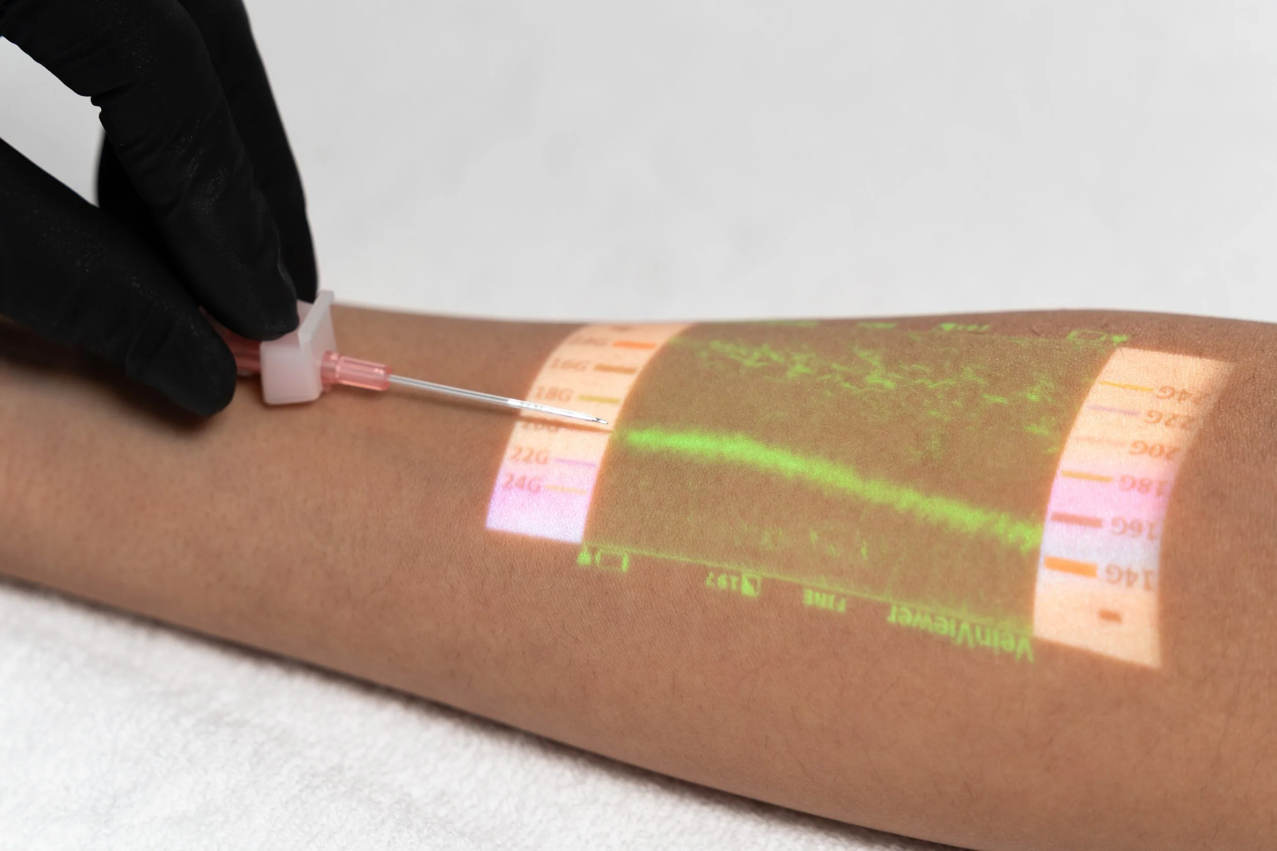
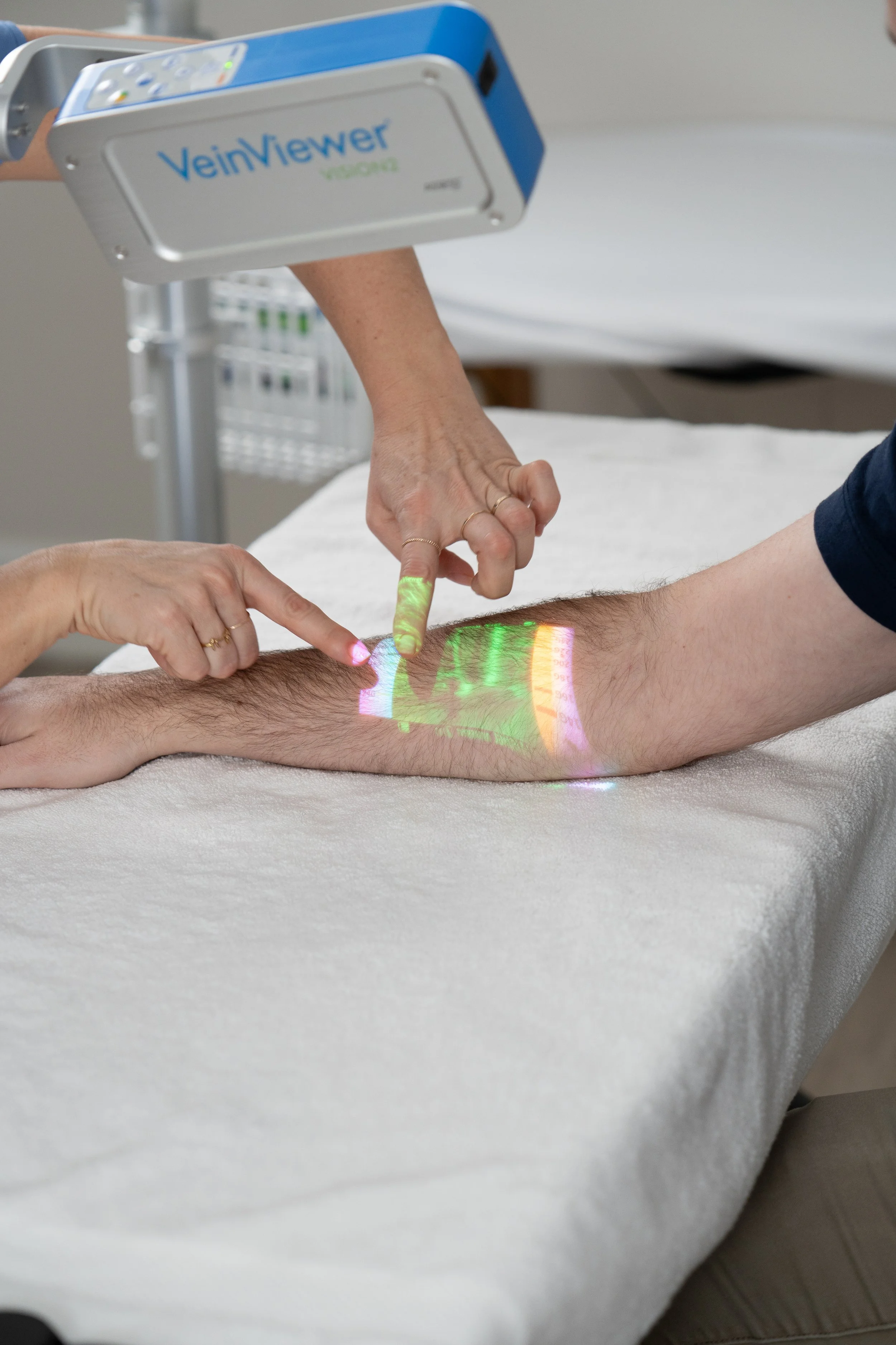
Available exclusively on VeinViewer® Flex and Vision2, CathCompass™ adds a built-in color-coded catheter sizing reference graphic to NIR vein visualization. Researchers studying PIVC outcomes, catheter dwell time, or vascular access complication rates can use CathCompass™ to standardize catheter selection based on vein size — a variable increasingly recognized in peripheral IV access literature.
When the appropriate catheter-to-vein ratio is selected, first-stick success increases to 92%.*
*van Loon F., et al., The impact of catheter to vein ratio on peripheral intravenous cannulation success, a post-hoc analysis; PloS One 2021; 16(5): e0252166Published Evidence Supporting VeinViewer®
VeinViewer® is backed by peer-reviewed research across multiple clinical settings, including:
Two Configurations for Research Settings
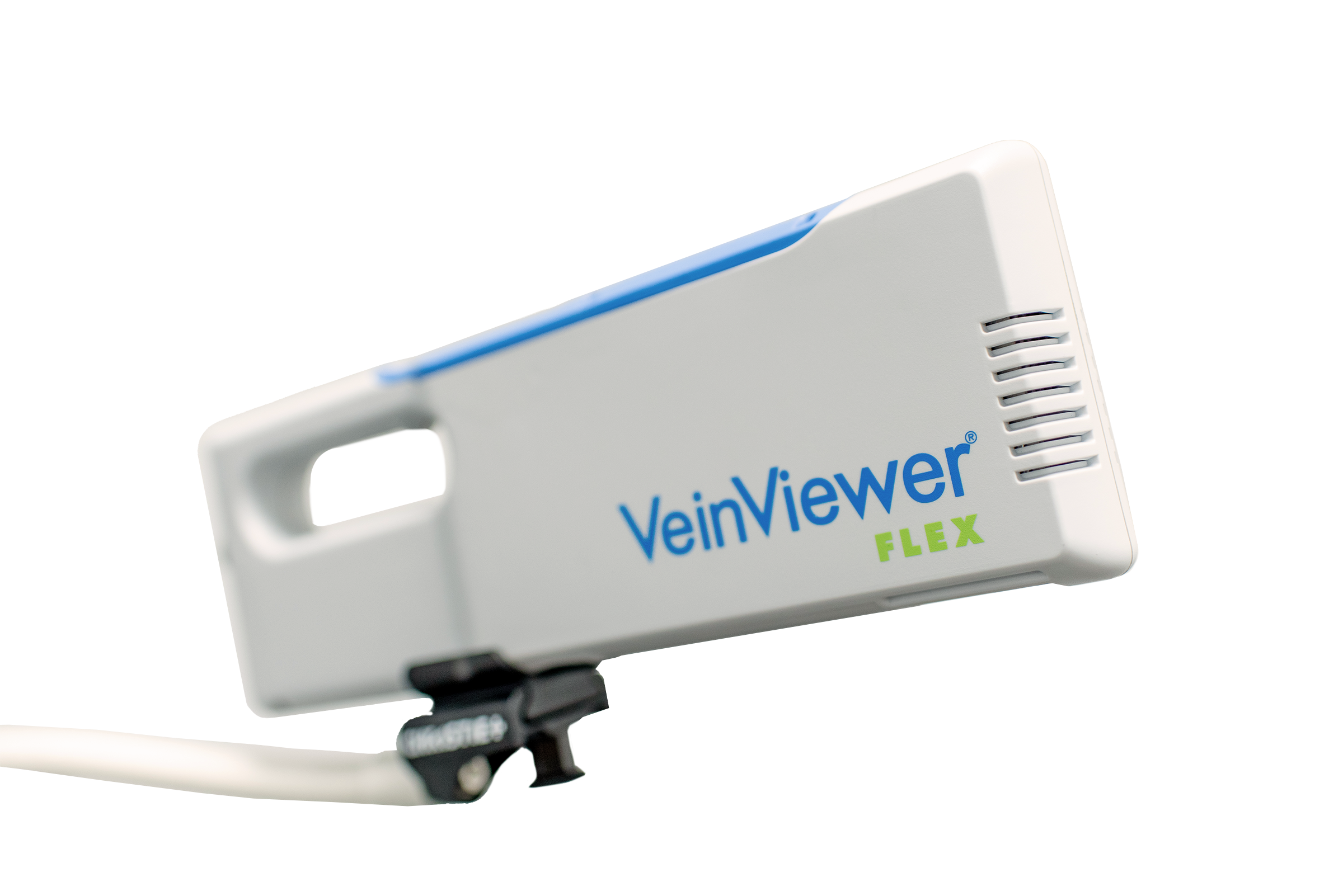
VeinViewer® Flex — Portable for Multi-Site and Field Research
Compact and lightweight, VeinViewer® Flex is ideal for multi-site studies, mobile research settings, veterinary labs, and clinical environments where portability matters. The VeinViewer® Flex is equipped with multiple imaging modes, including Fine Detail, Resize, MaxBright, and Inverse. The same core NIR imaging engine as Vision2, in a form factor that travels with your team.
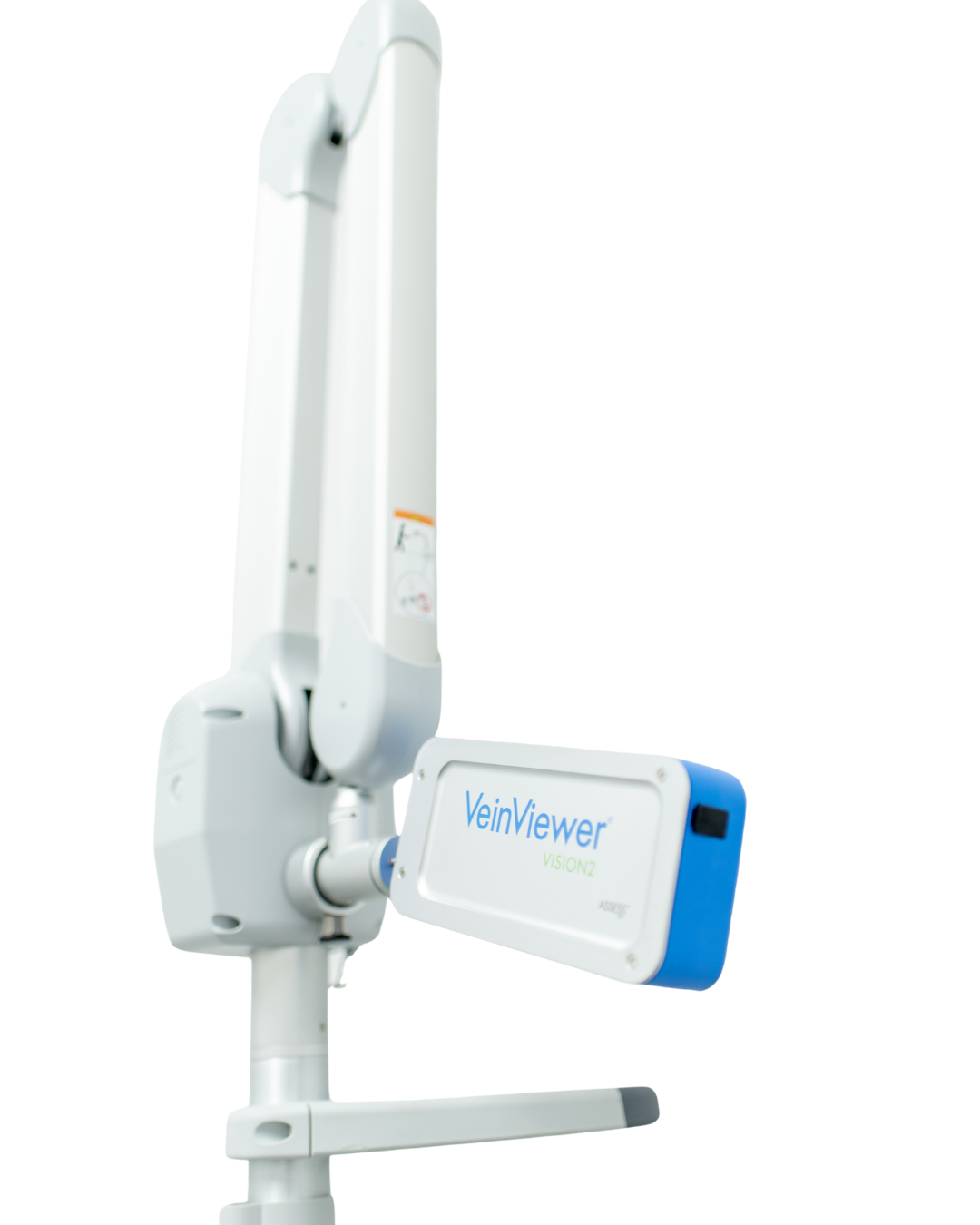
VeinViewer® Vision2 — Hands-Free for Controlled Clinical Settings
VeinViewer® Vision2 is designed for high-volume, controlled research environments. Its hands-free articulating arm, multiple imaging modes (TriColor, Fine Detail, Resize, MaxBright, Inverse), and image capture capability make it well-suited for procedural research, nursing education labs, and academic medical centers.
Bring VeinViewer® Into Your Research Program
Whether you’re conducting IRB-approved clinical trials, building a vascular access curriculum, or exploring NIR technology in a translational context, VeinViewer® is designed to support your goals.